
Go Optical
High-Performance Imaging for Stereo-seq Workflows
Go Optical
High-Performance Imaging for Stereo-seq Workflows
High Resolution
12-megapixel imaging enables accurate segmentation at tissue and single-cell level.
Large Travel Range
Motorized XY travel range of 100 mm x 70 mm, ensuring flexibility for diverse tissue types.
Fast Imaging
Scan a 10 mm x 10 mm chip in under 70 seconds using auto-focus and a 10x objective lens.
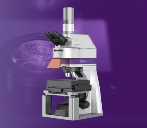
Go Optical is a STOmics microscope specifically designed to meet the imaging needs of Stereo-seq workflows. It employs multiple illumination techniques, including epifluorescence and transmitted bright-field microscopy, with DAPI, FITC, TRITC, and CY5 fluorescence channels. This advanced system enables efficient whole-tissue scanning and imaging, ensuring high-quality data acquisition for spatial biology applications.
Go Optical captures high-resolution images with minimal stitching errors, making it ideal for tissue segmentation in Stereo-seq applications. With a large scanning range, rapid imaging speed, clear image quality, and minimal stitching errors, it is the ideal choice for all Stereo-seq imaging applications. Equipped with an auto-focus mode, Go Optical can scan 10 mm x 10 mm chip within 70 seconds. By combining high-resolution imaging with advanced algorithms in the Stereo-seq analysis pipeline, it enables precise localization of gene expression on tissue sections.

High Resolution
12-megapixel super-resolution imaging accurately captures track lines and tissue details, enabling precise segmentation at both the tissue and single-cell levels.
Fast Imaging
With an unmatched imaging speed, the system scans a 10 mm × 10 mm chip in under 70 seconds using auto-focus mode and a 10X lens.

Large Travel Range
A motorized XY travel range of 100 mm × 70 mm accommodates the scanning needs of most STOmics Stereo-seq chips, ensuring flexibility for various sample sizes.

Applications
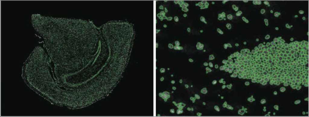
Go Optical imaging has various applications, including optimizing permeabilization conditions, mapping cell nuclei, and analyzing protein and tissue morphology through fluorescence photography for subsequent bioinformatics analysis.
Permeabilization Optimalization
Imaging can help determine the optimal permeabilization time by assessing the strength of tissue fluorescence signals across different chips processed at varying time points.

Spatial Mapping of Cell Nuclei
Fluorescence photography enables the precise recording of
cell nuclei positions and tissue morphology for downstream
bioinformatics analysis. This allows for high-resolution
visualization of cellular and tissue structures, facilitating
advanced techniques such as spatial mapping and
transcriptomic profiling.

Technical Specifications
| Go Optical | |
|---|---|
| Optical System | Positive scanning microscope |
| Objective Lens | 4X, 10X, 20X, 40X (optional) universal motorized objective lens converter, support expansion |
| Illumination Methods | Epi-fluorescence, Epi-bright field, Transmitted bright field |
| Fluorescent Channel | Standard configuration includes DAPI, FITC, TRITC, CY5 Optional configuration can support other dye filter blocks |
| Motorized Travel Range XY | 100 mm x 70 mm |
| Camera resolution | High image resolution: 3008 pixels (height) and 4112 pixels (width) |
| Auto-focus modes | Auto-focus mode and map navigation mode |
| Imaging stitching accuracy | ≤5 μm (10X) |
| Pixel size | 3.45 μm |
| Scanning Speed | ≤ 70 seconds for a 10 mm × 10 mm chip (10X) |
| Operating mode | Manual operation for human eye observation Fully automatic scanning and imaging |
Ordering Information
| Category | Product | Cat. No. |
|---|---|---|
| Instrument | STOmics Microscope- Go Optical | 900-001019-00 |
| Accessories | Objective Lens (40X) | 060-000217-00 |